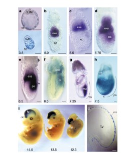
Eomesodermin is required for mouse trophoblast development and mesoderm formation
The earliest cell fate decision in the mammalian embryo separates the extra-embryonic trophoblast lineage, which forms the fetal portion of the placenta, from the embryonic cell lineages. The body plan of the embryo proper is established only later at gastrulation, when the pluripotent epiblast gives rise to the germ layers ectoderm, mesoderm and endoderm. Here we show that the T-box gene Eomesodermin 1 performs essential functions in both trophoblast development and gastrulation. Mouse embryos lacking Eomesodermin arrest at the blastocyst stage. Mutant trophoectoderm does not differentiate into trophoblast, indicating that Eomesodermin may be required for the development of trophoblast stem cells 2 . In the embryo proper, Eomesodermin is essential for mesoderm formation. Although the specification of the anterior–posterior axis and the initial response to mesoderm-inducing signals is intact in mutant epiblasts, the prospective mesodermal cells are not recruited into the primitive streak. Our results indicate that Eomesodermin defines a conserved molecular pathway controlling the morphogenetic movements of germ layer formation and has acquired a new function in mammals in the differentiation of trophoblast.
This is a preview of subscription content, access via your institution
Access options
Subscribe to this journal
Receive 51 print issues and online access
196,21 € per year
only 3,85 € per issue
Buy this article
- Purchase on SpringerLink
- Instant access to full article PDF
Prices may be subject to local taxes which are calculated during checkout


Lineage regulators TFAP2C and NR5A2 function as bipotency activators in totipotent embryos
Article 19 January 2024
The T-box transcription factor Eomesodermin governs haemogenic competence of yolk sac mesodermal progenitors
Article 08 January 2021
Initiation of a conserved trophectoderm program in human, cow and mouse embryos
Article 23 September 2020
References
- Ryan, K., Garrett, N., Mitchell, A. & Gurdon, J. B. Eomesodermin, a key early gene in Xenopus mesoderm differentiation. Cell87, 989–1000 (1996). ArticleCASPubMedGoogle Scholar
- Tanaka, S., Kunath, T., Hadjantonakis, A. K., Nagy, A. & Rossant, J. Promotion of trophoblast stem cell proliferation by FGF4. Science282, 2072 –2075 (1998). ArticleADSCASPubMedGoogle Scholar
- Herrmann, B. G., Labeit, S., Poustka, A., King, T. R. & Lehrach, H. Cloning of the T gene required in mesoderm formation in the mouse. Nature343, 617– 622 (1990). ArticleADSCASPubMedGoogle Scholar
- Papaioannou, V. E. & Silver, L. M. The T-box gene family. Bioessays20, 9– 19 (1998). ArticleCASPubMedGoogle Scholar
- Smith, J. C. T-Box genes: What they do and how they do it. Trends Genet.15, 154–158 (1999). ArticleCASPubMedGoogle Scholar
- Hancock, S. N., Agulnik, S. I., Silver, L. M. & Papaioannou, V. E. Mapping and expression analysis of the mouse ortholog of Xenopus eomesodermin. Mech. Dev.81, 205–208 (1999). ArticleCASPubMedGoogle Scholar
- Ciruna, B. G. & Rossant, J. Expression of the T-box gene eomesodermin during early mouse development. Mech. Dev.81, 199–203 (1999). ArticleCASPubMedGoogle Scholar
- Bulfone, A. et al. Expression pattern of the Tbr2 (Eomesodermin) gene during mouse and chick brain development. Mech. Dev.84, 133–138 (1999). ArticleCASPubMedGoogle Scholar
- Sutherland, A. E., Calarco, P. G. & Damsky, C. H. Expression and function of cell surface extracellular matrix receptors in mouse blastocyst attachment and outgrowth. J. Cell Biol.106, 1331–1348 (1988). ArticleCASPubMedGoogle Scholar
- Sutherland, A. E., Calarco, P. G. & Damsky, C. H. Developmental regulation of integrin expression at the time of implantation in the mouse embryo. Development119, 1175–1186 (1993). CASPubMedGoogle Scholar
- Rossant, J. & Spence, A. Chimeras and mosaics in mouse mutant analysis. Trends Genet.14, 358– 363 (1998). ArticleCASPubMedGoogle Scholar
- Lawson, K. A., Meneses, J. J. & Pedersen, R. A. Clonal analysis of epiblast fate during germ layer formation in the mouse embryo. Development113, 891–911 (1991). CASPubMedGoogle Scholar
- Thomas, P. & Beddington, R. Anterior primitive endoderm may be responsible for patterning the anterior neural plate in the mouse embryo. Curr. Biol.6, 1487–1496 (1996). CASPubMedGoogle Scholar
- Beddington, R. S. & Robertson, E. J. Anterior patterning in mouse. Trends Genet.14, 277 –284 (1998). ArticleCASPubMedGoogle Scholar
- Tada, M., Casey, E. S., Fairclough, L. & Smith, J. C. Bix1, a direct target of Xenopus T-box genes, causes formation of ventral mesoderm and endoderm. Development125, 3997–4006 (1998). CASPubMedGoogle Scholar
- Casey, E. S. et al. Bix4 is activated directly by VegT and mediates endoderm formation in Xenopus development. Development126, 4193–4200 (1999). CASPubMedGoogle Scholar
- Pearce, J. J. H. & Evans, M. J. Mml, a mouse Mix-like gene expressed in the primitive streak. Mech. Dev.87, 189–192 (1999). ArticleCASPubMedGoogle Scholar
- Rossant, J. & Tamura-Lis, W. Effect of culture conditions on diploid to giant-cell transformation in postimplantation mouse trophoblast. J. Embryol. Exp. Morphol.62, 217– 227 (1981). CASPubMedGoogle Scholar
- Feldman, B., Poueymirou, W., Papaioannou, V. E., DeChiara, T. M. & Goldfarb, M. Requirement of FGF-4 for postimplantation mouse development. Science267, 246–249 (1995). ArticleADSCASPubMedGoogle Scholar
- Arman, E., Haffner-Krausz, R., Chen, Y., Heath, J. K. & Lonai, P. Targeted disruption of fibroblast growth factor (FGF) receptor 2 suggests a role for FGF signaling in pregastrulation mammalian development. Proc. Natl Acad. Sci. USA95 , 5082–5087 (1998). ArticleADSCASPubMedPubMed CentralGoogle Scholar
- Liu, P. et al. Requirement for Wnt3 in vertebrate axis formation. Nature Genet.22, 361–365 (1999). ArticleCASPubMedGoogle Scholar
- Ding, J. et al. Cripto is required for correct orientation of the anterior–posterior axis in the mouse embryo. Nature395, 702 –707 (1998). ArticleADSCASPubMedGoogle Scholar
- Tam, P. P. & Behringer, R. R. Mouse gastrulation: the formation of a mammalian body plan. Mech. Dev.68, 3–25 (1997). ArticleCASPubMedGoogle Scholar
- Wilson, V., Manson, L., Skarnes, W. C. & Beddington, R. S. The T gene is necessary for normal mesodermal morphogenetic cell movements during gastrulation. Development121, 877 –886 (1995). CASPubMedGoogle Scholar
- Wilson, V., Rashbass, P. & Beddington, R. S. Chimeric analysis of T (Brachyury) gene function. Development117, 1321– 1331 (1993). CASPubMedGoogle Scholar
- Ciruna, B. G., Schwartz, L., Harpal, K., Yamaguchi, T. P. & Rossant, J. Chimeric analysis of fibroblast growth factor receptor-1 (Fgfr1) function: a role for FGFR1 in morphogenetic movement through the primitive streak. Development124, 2829– 2841 (1997). CASPubMedGoogle Scholar
- Sun, X., Meyers, E. N., Lewandoski, M. & Martin, G. R. Targeted disruption of Fgf8 causes failure of cell migration in the gastrulating mouse embryo. Genes Dev.13, 1834– 1846 (1999). ArticleCASPubMedPubMed CentralGoogle Scholar
- Wattler, S., Russ, A., Evans, M. & Nehls, M. A combined analysis of genomic and primary protein structure defines the phylogenetic relationship of new members of the T-box family. Genomics48, 24–33 (1998). ArticleCASPubMedGoogle Scholar
- Hogan, B., Beddington, R., Constantini, F. & Lacy, E. Manipulating the Mouse Embryo (Cold Spring Harbor Laboratory Press, New York, 1994). Google Scholar
Acknowledgements
We thank J. Gurdon, A. Bulfone, A. Zorn, N. Papalopulu, D. St. Johnston and R. Pedersen for discussion; X. Sun and F. Beck for communicating results before publication; G. Martin, C. Wright and R. Milner for gifts of probes and reagents; F. Wianny and A. Sossick for help with confocal microscopy; J. Wilson for technical assistance; J. Ferguson, P. Whiting and R. Plumridge for animal care. A.P.R. thanks W. Gross for continuing support and advice. This work was funded by grants from the Wellcome Trust. V.W. is supported by a Medical Research Council Career Development Award.
Author information
- Sigrid Wattler & Michael C. Nehls Present address: Ingenium Pharmaceuticals AG, Lochhamer Strasse 29, 82152, Martinsried, Germany
- Kenneth Ryan Present address: Children's Hospital of Philadelphia, University of Pennsylvania School of Medicine, 34th and Civic Center Blvd, Philadelphia, PA 19104, USA
- Martin J. Evans Present address: Cardiff School of Biosciences, Museum Avenue, PO Box 911, Cardiff, CF10 3US, Wales, UK
Authors and Affiliations
- Wellcome/CRC Institute for Cancer and Developmental Biology, University of Cambridge, Tennis Court Road, Cambridge, CB2 1QR, UK Andreas P. Russ, Samuel A. J. R. Aparicio, Mark B. L. Carlton, Jonathan J. Pearce, Sheila C. Barton, M. Azim Surani, Kenneth Ryan & Martin J. Evans
- Department of Physiology, University of Cambridge, Downing Street, Cambridge, CB2 3EG, UK Andreas P. Russ, William H. Colledge & Mark B. L. Carlton
- Paradigm Therapeutics Ltd, Downing Street, Cambridge, CB2 3EG, UK Andreas P. Russ, Samuel A. J. R. Aparicio & Mark B. L. Carlton
- Niedersächsisches Institut für Peptidforschung, Feodor-Lynen-Strasse 31, Hannover, 30625 , Germany Sigrid Wattler & Michael C. Nehls
- Dep. of Oncology, Cambridge Institute of Medical Research, Addenbrookes' Hospital, Cambridge, CB2 2XY, UK Samuel A. J. R. Aparicio
- Centre of Genome Research, University of Edinburgh, Kings Buildings, West Mains Road, Edinburgh, EH9 3JQ, UK Valerie Wilson
- Andreas P. Russ
